coral morphologic header


Archive for the ‘Research’ Category

Lower Keys Coral Bleaching Report (August 22, 2014)

Monday, August 25th, 2014

Having been preoccupied with the Miami Coral Rescue Mission this summer, we finally made our first excursion to the Lower Keys this summer on Friday August 22. Sadly, we found that a distressingly high percentage of corals living on the reefs in Hawk Channel are severely bleached. Most of the staghorn corals that we saw were severely bleached or actively dying, though there were a few hardy exceptions. Nearly all of the brain corals were bone white. All over the reef we observed an unhealthy mix of cyanobacteria and algae proliferating on previously dead coral skeletons. Even the normally hardy gorgonians, corallimorphs, and zoanthids showed significant bleaching on all three patch reefs we checked. The water temperature was an uncomfortable 89 degrees on the bottom. Without strong winds or storms to cool off the water, we are concerned that many reefs in the Keys will lose significant coral cover in the next several months.

(more…)

Miami Coral Rescue Retrospective & Urban Coral Hypothesis

Monday, June 9th, 2014

Meandrina meandrites Juvenile 1 SM

A hyper-fluorescent juvenile Montastrea cavernosa rescued from Government Cut.

After months of impatiently watching dredge ships working offshore Miami, Coral Morphologic and other researchers were finally granted a brief window of opportunity from May 26 until June 6th in which to rescue corals left behind from the legally-required relocation effort from the Army Corps of Engineers’ Deep Dredge of Government Cut. This was a much shorter length of time than we had been prepared for, and as such, we had to respond with considerable urgency in order to rescue as many corals as possible. Fortunately we had begun our detailed preparations in January 2014 by coordinating students and professors from the University of Miami to help in the effort. Collectively, the Miami Coral Rescue Mission removed over 2,000 stony corals that would have otherwise been destroyed in the process to make way for the larger ships that will pass through the soon-to-be-expanded Panama Canal.

The majority of the corals that Coral Morphologic removed from Government Cut have now been transplanted to an artificial reef about one mile south from where they originated, and where we will continue to monitor them to ensure their long-term survival. Some corals will be sent to the Smithsonian Institution for research. And the rest of the corals were brought back to our Lab, where we will document them via film and photography for a body of work titled ‘Coral City’, in which we will present them as fluorescent icons for a 21st century Miami.

While we could have rescued more corals with an extended deadline, the Miami Coral Rescue Mission is not over. It is now entering a longer-term monitoring phase in which we will continue to assess the health of surrounding coral reefs through July 2015, when the Deep Dredge project is finally slated for completion.

Read a New York Times article on the rescue mission and listen to an NPR story below covering a day out on the water. Click the link below the NPR story to read the remainder of this post.

(more…)

‘Species Diversity of Shallow Water Zoanthids (Cnidaria: Anthozoa: Hexacorallia) in Florida’

Thursday, October 4th, 2012

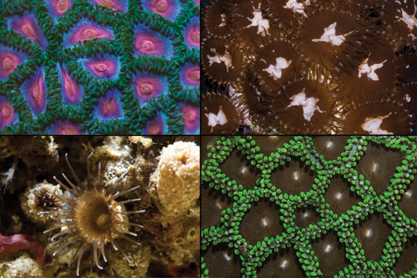
Four Floridian zoanthids analyzed in our study. Clockwise from Top Left: 1) Undescribed Zoanthus aff. pulchellus 2) Undescribed Palythoa aff. variabilis 3) Zoanthus solanderi 4) Undescribed Terrazoanthus sp.

Recently, we spearheaded a study of the Zoanthids found in our local nearshore waters that has been published in the Journal of Marine Sciences titled ‘Species Diversity of Shallow Water Zoanthids (Cnidaria: Anthozoa: Hexacorallia) in Florida‘ with Dr. James Reimer and Yuka Irei of the University of the Ryukyus in Okinawa, Japan. This is the first comprehensive study of its kind, analyzing DNA to determine the taxonomic authenticity of our local zoanthid species. We discovered that there are as many as four species of zoanthids in South Florida that have been overlooked by scientists until now.

Despite their ubiquitousness in shallow tropical waters, zoanthids have been largely neglected by marine biologists who have otherwise been more focused on understanding reef-building stony corals, leaving the taxonomy of tropical zoanthids vague and out of date. This, combined with the natural morphologic variability of these animals, makes physical identification difficult for the casual observer. The advent of DNA analysis has allowed for an accurate picture to emerge, and it is clear that there is much more diversity than had previously been recorded.

Read ‘Species Diversity of Shallow Water Zoanthids (Cnidaria: Anthozoa: Hexacorallia) in Florida‘:

Colins-Zoanthid-Paper-2012

‘Anemone Spawn’

Thursday, May 31st, 2012

A fluorescent green flower anemone (Epicystis crucifer) releases sperm into the water column at the Coral Morphologic lab.

On May 24th we observed this fluorescent green flower anemone (Epicystis crucifer) spawning in our lab, and managed to film the event. The anemone continued to release sperm for nearly 30 minutes, while several other nearby flower anemones released significantly smaller amounts of gametes. This was the second time we have witnessed a flower anemone spawning event at our lab this spring. We first observed a synchronous spawn of more than a dozen anemones in an outdoor aquaculture system that receives natural sunlight on April 12th. After the jump are photos of anemones spawning during this event.

(more…)

‘(How To Grow) A Floating Forest’

Monday, October 3rd, 2011

In order to understand what’s going on in the video, you’re going to want to read the post below!

One of the most innovative, practical, and functional coral nurseries on the planet can be found just a few miles off the shores of Key Largo. The nursery consists of thousands of neatly organized colonies of the critically important staghorn coral (Acropora cervicornis) grown by the Coral Restoration Foundation (CRF) for the purpose of transplantation back to the reef. Staghorn corals have been decimated by disease and extreme weather here in Florida over the past 30 years, resulting in a seriously degraded reef ecosystem. Fortunately the CRF has developed methods that maximize the growth potential of these corals in their nursery, demonstrating that coral aquaculture is a realistic and effective way to restore beleaguered wild populations.

(more…)

‘Unidentified Ricordea Shrimp’

Monday, January 4th, 2010

‘Unidentified Ricordea Shrimp’
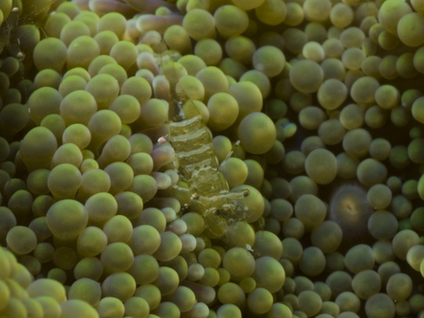
Unidentified commensal shrimp on Ricordea florida corallimorphs
Music, Video, and Aquarium
2010 Coral Morphologic

Shown above is the first documented video of a currently unidentified shrimp commensal with Ricordea florida corallimorphs. The nearly invisible shrimp measures only 9mm in total length. The ricordea polyp is about 30mm in diameter for comparison. We first reported and photographed this shrimp in October 2009. Subsequently, we sent a preserved specimen to taxonomist Dr. Richard Heard at the Gulf Coast Research Laboratory. Dr. Heard was unable to match the specimen with a described shrimp species. We will be sending additional specimens in the near future to confirm this shrimp’s newness to science.

Unidentified Caribbean Palythoa sp.

Friday, November 13th, 2009

Unidentified Palythoa sp.

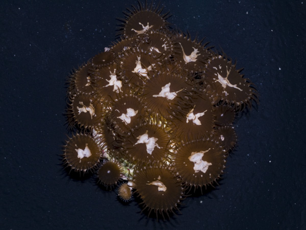
Unidentified ‘Butterfly’ Palthoas.

Recently while diving off of Key West, I was fortunate to come upon a rare and unidentified species of Palythoa. This was the first time that I have come upon this type in five years of frequent diving throughout the Florida Keys. Apparently it is less rare elsewhere in the Caribbean, but as of now has yet to be properly identified by a zoanthid taxonomist. On its particular patch of reef it was relatively abundant, despite being completely absent in seemingly identical reefs in the surrounding area. And while it might seem logical that it only takes one lucky zoanthid larvae to ultimately colonize a large area, it seems that there were at least 2 separate morphs cohabiting the area, which makes its complete absence from other nearby reefs compelling. And while Caribbean Palythoa display morphologies that seem to overlap, these particular Palythoa have a few traits that make it noticeably distinct:

  • Small size (1/4″-3/8″ disc diameter)
  • Translucent oral disc, often with teal-bluish iridescent sheen
  • Distinctive white splotch, often butterfly shaped
  • Eyelash-like tentacles

But considering that the morphologies of Caribbean Palythoa species seem to blur together, genetic analysis will be the most reliable way to determine species-hood. Fortunately our friend James Reimer is a zoanthid expert in Japan’s Ryuku Islands with access to such equipment and expertise. In addition to sending him samples of this Palythoa morph, we will also include a variety of other local Palythoa morphs to see just how distinct the individual species are.

Unidentified Commensal Ricordea Shrimp

Monday, October 19th, 2009

Unidentified Ricordea Shrimp

Pictured above is a very tiny (10mm) shrimp that lives commensally with Ricordea florida polyps.

Over the past several years I have occasionally encountered fleeting glimpses of tiny shrimp that live amongst the pseudo-tentacles of Ricordea florida. On all the previous occasions that encountered one, I had never been properly equipped with a super-macro camera  kit. A dive this past September finally warranted a good photo. Ricordea shrimp are tiny (8-12mm) and nearly transparent, making them very difficult to detect. It is unlikely that these might be juveniles of a more common commensal species (e.g. Periclimenes pedersoni, P. rathbunae, or P. yucatanicus), as it is clear from the photo above (and from recently collected specimens) that they are mature egg-bearing females at this small size. If you look closely at the photo, you’ll notice in the upper left-hand side of the photo that there is another pair of eyes. At the time of the photo I didn’t notice that there were several other tiny and completely clear shrimp living with this female. It was only while viewing the photos close-up that I noticed these other shrimp. Most likely they are male or juvenile females living colonially. On subsequent dive trips I have found several groups (3-5) of these tiny shrimp all living on the same R. florida colony.

Today we are sending off a preserved specimen to Periclimines shrimp expert Dr. Stephen Spotte for taxonomic inspection. He will be able to determine whether this shrimp has been previously identified, or whether we are dealing with a new species altogether.

Aberrant Tissue Inflation of Diploria clivosa

Friday, July 10th, 2009

July 9, 2009

The brain coral (Diploria clivosa) colony pictured above featured several areas ranging in size from 3-6 cm that exhibited very unusual cauliflower-like tissue expansion with warty protuberances. The photo was taken offshore of South Beach, Miami, Florida.

July 7, 2009

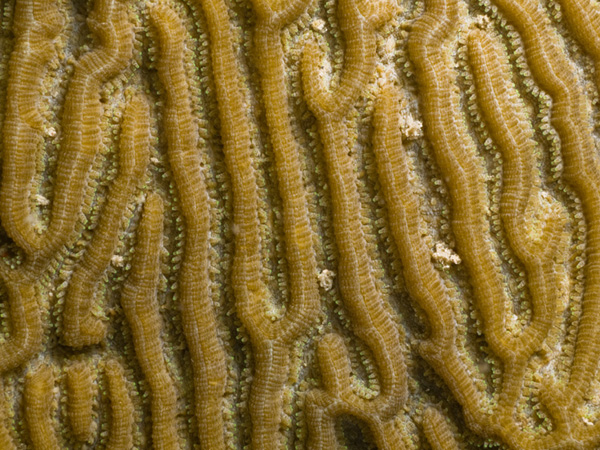
Pictured above is the normal ‘meandroid’ growth form for the brain coral Diploria clivosa. The tissue is relatively compact against the skeleton and the tentacles are visible along the inner walls of the grooves.

Summer Solstice Birthing

Monday, July 6th, 2009

Baby flower anemones attached near the base of "momma"

Two new-born Epicystis crucifer anemones are attached to Valonia sp. bubble algae at the base of their “mother” anemone.

On the evening before the summer solstice, we noticed that several of our favorite flower anemones (Epicystis crucifer) were exhibiting classic signs of stress (gaping mouth, regurgitation, decreased turgor pressure). However, all the water parameters suggested nothing out of the ordinary, so we simply decided to leave them alone, while monitoring them closely. The following morning we began finding tiny flower anemones attached to the aquarium bottom nearest to  the anemones that had appeared stressed. By this time though, the adult flower anemones had returned to their normal, healthy condition as if nothing had occurred. It was clear that the ‘stress’ that they were going through was a precursor to birthing dozens of 2mm babies from their mouths. As you can see from the photos, these babies are practically fully-formed, miniature versions of the adults.

To our knowledge, this is the first documented case of Epicystis crucifer birthing in a captive (or wild) environment. Live birth is not uncommon for many other anemone species, and given the local abundance of this species, it makes sense. It still remains unclear whether these babies were brooded internally from sexual reproduction, or whether they are asexual clones. Every little piece of information that we can gather on the life cycle of these beautiful animals is an important advance.

(more…)

Aberrant Tentacles of Ricordea florida

Saturday, April 18th, 2009

aberrant tentacles of a Ricordea florida polyp

This neon green Ricordea florida polyp displays a few abnormally large tentacles. This is an unusual characteristic not yet observed before in the Coral Morphologic Lab.

Fluorescent Pycnogonid (Sea Spider)

Tuesday, April 7th, 2009

Morphologic Studios 2009 Colin Foord

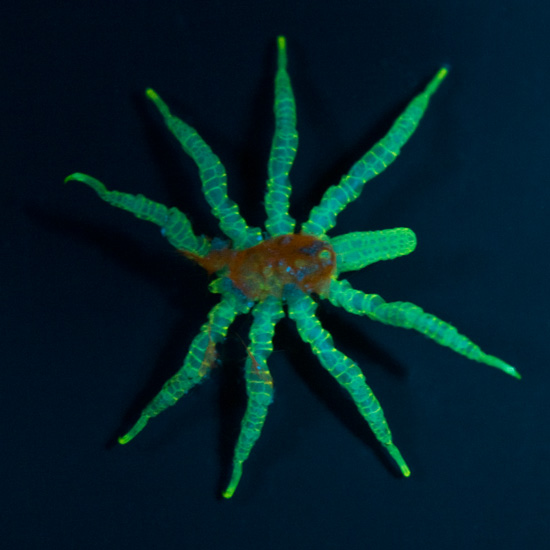
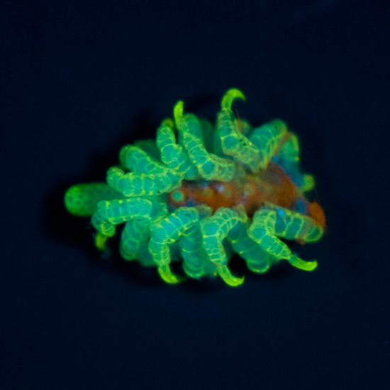
The cephalothorax of this unidentified pycnogonid is covered in wisps of  cyanobacteria (perhaps as camouflage) which itself fluoresces orange-red. This specimen measures about 1 cm in diameter. Fluorescence photograph.

It is amazing the wide variety of sea creatures that demonstrate fluorescence. Animals that you would never suspect to “glow” do just that when illuminated with the proper wavelength light. For the past several years I have been using an underwater flashlight that I custom outfitted with 470nm blue LEDs in order to be able to scan for glowing creatures in the darkness. This pycnogonid sea spider (not an arachnid) is a prime example of an unexpected life form displaying an ability to fluoresce. The “spider web”-like pattern of fluorescence over this specimen is truly impressive.

Pycnogonids, commonly known as “sea spiders” are rarely seen in the wild (but not necessarily rare), and are an unusual class of arthropod. Most are very small (<1cm), and hence overlooked. However, in the Antarctic there are some monsters that can grow up to 90cm (Pycnogonaphobia?)! One of their most distinctive features is that they possess a proboscis (a drinking straw if you will) that allows them to suck out fluids from soft-bodied invertebrates such as corals, anemones, and corallimorphs. In the wild they rarely do lasting damage to their prey. However within the confines of an aquarium I could imagine that this might not be the case where their selection of hosts is limited. As such, they should be regarded as potential parasites of corals within the aquarium, and removed if detected. Despite this threat, they don’t seem to be widespread pests within the aquarium keeping world.

Morphologic Studios 2009 Colin Foord

The same pycnogonid specimen in a defensive posture after being prodded “into position” for photography.

Coral Mucus and Bacteria: A Symbiosis?

Monday, July 14th, 2008

Anyone who has ever scraped or cut their skin on a living coral can attest to the malignant nature of what should otherwise be a minor abrasion or cut. These scrapes don’t heal very quickly, and can become infected very easily. The reason is that the mucus coating secreted by the coral harbors a dense population of bacteria that apparently gains protection and nutrition from said mucous. The relationship between mucous bacteria and coral is only now beginning to be unraveled.

That there were at least 16 presentations and posters that focused specifically on coral mucus at the ICRS, shows the level of interest this topic has been receiving within the marine biological world.

Several of the research projects concluded that the bacterial populations within the coral mucus are in fact mostly unique and independent of the bacterial populations that are found in nearby environments (surface sediments, biofilms, water column, etc). This indicates that the coral mucus/ bacteria relationship is more complex and specific than previously thought.

An important research topic on coral mucus bacteria revolves around their relationship to coral immune health and disease prevention. It is speculated that the mucus bacteria are somehow capable of thwarting coral infections (other bacteria, protozoans, etc), by maintaining a balanced population within the mucus (perhaps a similar theory of using ‘probiotics’ as preventative measure?). Whatever the mechanism, it appears that the coral mucus and resident bacteria population acts as a protective barrier against pathogenic invaders.

Another important role of coral mucus is to act as a medium for nutrient transport. I assume that this perhaps relates to the mucus’ ability to help adhere to and coat food particles, thereby aiding and enabling digestion. This is a conjecture on my part, but it seems like a logical process.

A research poster entitled “A Quantitative Approach Linking Coral Mucus And Their Symbiotic Zooxanthellae in Response To Environmental Change” found that 45% of the daily fixed carbon (i.e. the food produced from photosynthesis), was incorporated into coral mucus in Montastrea annularis. This demonstrates the vital importance that coral polyps place on mucus production.

In the poster mentioned above, the researchers determined that as water temperatures were increased by 1.5 degrees C, mucocyte density (specialized cells that produce mucus) increased, while zooxanthellae density decreased. They draw the conclusion that increasing temperatures cause M. annularis to rely more upon heterotrophy (eating), than upon autotrophy (zooxanthellate photosynthesis). Bleached corals were found to have lower densities of mucocytes, but the remaining mucocytes were greatly enlarged, indicative of highly increased mucus production per mucocyte.

A thought that popped into my head while reading the results of this paper, in combination with the other information I picked up in several lectures on the topic, is that perhaps the coral actually digests the bacteria that live in the mucus layer, thereby adding an additional symbiotic food source (Zooxanthellae being the other “food” producer). It seems possible that by providing a suitable medium for bacterial growth, the coral is able to culture its own “bacteria garden” that is consumed at a rate that is balanced with mucus production and the bacterial growth within it.

I hope that future research continues to look into this area, as it is possible that there is still a piece of the coral nutrition puzzle that is still waiting to be unraveled.

Corallimorpharians, etc. @ ICRS

Friday, July 11th, 2008

There has been recent interest in the corallimorpharians by marine biologists, due to genetic studies that demonstrate that some hard corals (Scleractinia) are actually more closely related to corallimorphs than to other members of Scleractinia. This has resulted in several papers that suggest that corallimorphs once had a Scleractinian ancestor with a calcium carbonate skeleton. Prior to these genetic studies, it had been suggested (through taxonomic studies) that corallimorphs were in fact more closely related to sea anemones (Actiniaria).

To explain why the loss of this skeleton may have occurred, researchers have been suggesting that increased levels of carbon dioxide during the Triassic period (from about 200-240 million years ago), would have caused a reduction in oceanic pH, which resulted in a decreased ability by hard corals to build a calcium carbonate skeleton. These conditions would therefore select for corals that could adapt to live without their skeletons. And voila!, the corallimorpharians were born. Unfortunately, the fossil record of corallimorpharians is minimal due to their lack of calcium carbonate skeleton, making this claim hard to prove. It is certainly a very seductive argument, especially now that atmospheric levels of CO2 are once again rising to levels that may soon have a detrimental impact on hard corals and their ability to continue reef building. This has some scientists positing that in the future as hard corals start dying off, soft bodied anthozoans will once again have an adaptive advantage, resulting in their proliferation.

An interesting poster entitled “Mechanisms of Microhabitat Segregation among Corallimorpharians”, examined the ability of two common Red Sea corallimorphs to handle UV light stress. One species Rhodactis rhodostoma, is commonly found in shallow-water reef flats where light intensity is very high. The other, Discosoma unguja, is common at deeper depths in shaded areas. The researchers exposed both species to identical conditions of light with several variable treatments (high light, shaded, etc.).

To understand what the researchers were looking for, as far as “light stress” is concerned, one must understand what happens within the corals’ tissues and zooxanthellae during photosynthesis. To summarize, one byproduct of zooxanthellae photosynthesis is the formation of highly reactive and potentially damaging oxygen ions. The coral must deactivate these harmful byproducts in order to prevent tissue damage. Fortunately, corals have evolved certain enzymes that are capable of deactivating these oxygen radicals. This is the reason why it is very easy to “light shock” your corals by rapidly increasing the amount of light they receive. The coral is simply unprepared to deactivate all of the additional harmful byproducts of the increased photosynthetic activity.   typical emergency reaction by the coral is to expel its zooxanthellae  (bleaching) so that no further damage can occur.

Anyway, back to the study at hand. When the Discosoma unguja were placed under full strength light, they showed a decided inability to handle the affects of increased photosynthetic activity. Instead of an enzymatic ability to deal with high light, they expressed an ability to move away from the light source. Crafty little creatures.

Rhodactis rhodostoma, on the other hand, proved to be resilient to high levels of light. Tissue analysis showed much higher levels of the enzymes necessary for deactivating harmful oxygen ions than in D. unguja.

What the study did not mention however, was whether the Discosoma unguja could be acclimated to higher light over a long period of time. And if so, would they be capable of gradually increasing their enzymatic abilities? If the Discosoma were taken from a shaded location and immediately exposed to high light, one could only expect a rapid stress response.   But as we aquarists have learned, corals can be highly adaptive to unnatural conditions so long as the conditions do not rapidly fluctuate.

This study seems to reinforce the hypothesis that as climactic and atmospheric conditions become more extreme, the corallimorphs are poised to be able to adapt (some might even say morph) swimmingly to changing water chemistry, nutrient and temperature increases, even as hard corals decline.

ICRS (Day 3)

Wednesday, July 9th, 2008

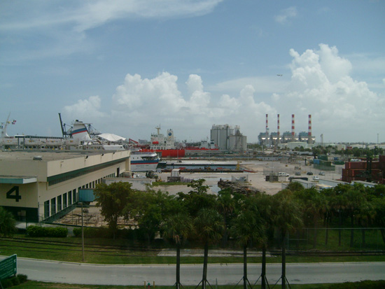
A view from the ICRS Fort Lauderdale. Cruise ships, smoke stacks, palm trees, cargo containers, jets, and blue skies… Welcome to Port Everglades.

Once again, an impressive number of topics relevant to reef aquarists on the third day of the International Coral Reef Symposium. To summarize:

In a lecture on the genetic diversity of Heliofungia actinoformis populations in Indonesia, I learned that the global trade in live corals is worth between 200-330 million US dollars annually (and no doubt increasing). This represents between 11-12 million corals exported each year. That’s a lot of coral.

____________________________________________________________________

In an in-situ (lagoon based) coral aquaculture experiment in the Maldives, researchers concluded that the most efficient way to maximize the development of axial polyps (fast growing branch tip polyps) in Acropora muricata (a “staghorn” species) was to do the following:

  • Use basal fragments (mid-branch, not branch tips, i.e. broken on both ends) laid down horizontally on the grow-out tray (perpendicular to the water surface).
  • Induce injury (by scraping off sections of basal polyps) at several locations along the top of the fragment to encourage the development of axial polyps that will form new branches.
  • The smallest fragment sizes in the experiment (3cm) maximized axial polyp formation. Productivity was increased by 75% by using three 3cm frags instead of one 9 cm frag.

It should be noted that the experiment took place in pristine water conditions, so survivability was nearly 100%. Algal overgrowth and disease was not an issue. I would expect that if this experiment took place in more nutrient laden water, that survivability would be reduced when inducing injury on such small fragments (3 cm). Nevertheless, I would have predicted the faster growth from branch tips rather than mid-section fragments, but in fact this sort of “pruning” clearly encourages the growth of multiple, fast-growing branches.

___________________________________________________________________

There was another interesting lecture entitled “Morphological Dependance of the Variation in the Light Amplification Capacity of the Coral Skeleton” that focused on the ability of a coral’s aragonite skeleton to amplify light by scattering it over a wider surface area, and ultimately providing surrounding polyps with more available light for photosynthesis.

The researchers tested different skeletal morphologies (i.e. massive, branching, plating, etc), and discovered that there was a clear correlation in light scattering depending on the corals’ growth form. For instance, the encrusting coral Porites branneri was able to absorb four times as much light over the same surface area as that of a smooth sea grass blade. Of all the corals tested Echinopora horrida (a branching coral) absorbed the most amount of light, where as Caulastrea furcata (phecelloid morphology i.e. polyps are separated with no connecting tissue) absorbed the least. It should be noted that Acropora branching corals scattered light so well that their lab equipment could not properly calculate the efficiency of this scattering. They estimate that Acropora is capable of absorbing more than 10 times the amount of light than what would otherwise hit a flat, non-aragonite surface. To summarize, they conclude the efficiency of light absorption more or less follows this morphological pattern:

(least) Solitary polyp < Phaceloid < Massive < Plating < Branching (Most)

___________________________________________________________________

A genetic analysis of the yellow tang (Zebrasoma flavescens) seemed to indicate the possibility that this species developed within the Hawaiian Archipelago (where it is common), and then more recently spreading south and west through the northern tropical/sub-tropical Pacific. Over this large geographic distance genetic variability was quite low, indicating wide larval dispersal carried by oceanic currents. Peculiarly, around the Big Island of Hawaii, up to four distinct genetic populations existed in rather close (but clearly separate) vicinities. It was posited that the yellow tang has not reached the Southern Indo-Pacific due to niche overlap and competition with the common scopas tang (Zebrasoma scopas). Where these two species occur at the same location, hybridization is common.

___________________________________________________________________

I got to spend some time with Brian Plankis and Eric Borneman to discuss their Project DIBS and Reef Stewardship Foundation. I will do my best to highlight these endeavors in a separate post. In the meantime check out their websites, and sign-up with Project DIBS (Desirable invertebrate Breeding Society).
Zoanthids @ ICRS (Day 2)

Tuesday, July 8th, 2008

One of the first presentations that I caught today at the ICRS focused on the genetic analysis of Indo-Pacific and Caribbean zoanthid species. The work was performed and presented by Dr. James Reimer, a zoanthid specialist at the University of the Ryukus in Japan. If you’ve ever spent any time on CoralPedia.com (formally Zoaid.com), you might be familiar with Dr. Reimer and his work. Take a look here to see his photo gallery.

For Indo-Pacific species he analyzed Zoanthus vietnamensis and Z. sansibaricus. For Caribbean species he analyzed Z. sociatus and Z. pulchellus. His findings revealed that Z. vietnamensis and Z. sociatus were genetically similar enough to be considered part of the same closely related clade (a taxonomic group with a common ancestor). Similarly, Z. sansibaricus and Z. pulchellus were shown to be grouped together in another clade. This suggests that at one time in the Earth’s history, these two pairs of zoanthid species were at one time the same species (Z. vietnamensis= Z. sociatus, Z. sansibaricus= Z. pulchellus). The separation of the species occurred when the isthmus of Panama closed about 3 million years ago.

Further genetic analysis of the Symbiodinium sp. (zooxanthellae) from Z. vietnamesis and Z. sociatus showed that these two species share identical symbiotic zooxanthellae, despite at least 3 million years of geographic isolation in different oceans. The same was true when he compared the zooxanthellae from Z. sansibaricus and Z. pulchellus. Dr. Reimer believes that the separation and development of the current species occurred about 6.5-7 million years ago.

Additionally, the two clades are similar enough with each other that genetic similarity suggests that all four current species shared a single common ancestor about 15 million years ago.

I asked Dr. Reimer if he had had a chance to compare the DNA from Caribbean and Pacific specimens of Zoanthus gigantus (known in the hobby as “People Eaters”). Unfortunately, he only has one sample of Caribbean Z. gigantus, and therefore hasn’t been able to do the appropriate analysis. To overcome this hurdle, we have offered to help supply him with these and other Caribbean zoanthids for future studies.

International Coral Reef Symposium (Day 1)

Monday, July 7th, 2008

Today began the first day of the 11th International Coral Reef Symposium (ICRS) in Fort Lauderdale, FL. These symposiums take place every four years (2004 was in Okinawa, 2000 was Bali), so we were incredibly lucky to have this world-class scientific event taking place right up the road from Miami. I will do my best to summarize the facts and insights that I think reef aquarists would find most interesting.

– The fungiid coral Ctenactis echinata and to a lesser extent Fungia repanda can engage in hermaphodism (sex change) over the course of their lifespans. Some can even change sex more than once (m->f->m). Apparently the smaller, younger individuals are more likely to be male, and as they get larger and can afford to invest more energy into egg production, they switch to female. At medium sizes, they can change sexes on alternate spawning years. Spawning takes place at 8:45 pm 5 nights after the full moon in July (at least in Japan where the research took place).

– While much attention has been made over the precipitous decline of Diadema urchins on Caribbean reefs, and the resulting overgrowth of macroalgae in the absence of said urchins, a study done in Panama suggests that urchins of the genus Echinometra are at least comparably effective at macroalgae removal as Diadema. Overgrowth from macroalgae is considered a primary roadblock to proper coral larvae settlement, and therefore herbivores like urchins are necessary to keep the coral reef ecosystem in balance.

– It was discovered by accident during an experiment on coral larvae settlement that the larvae of Porites asteroides showed a decided preference for settlement on fluorescent orange/red plastic cable ties. In fact, they settled significantly more so on these cable ties than on the expected preferred substrate of crustose coralline algae (similarly pink/purple in color). The researchers then investigated the role of the chemical rhodopsin in the selection of substrate. Rhodopsin is the chemical found in the human eye that absorbs blue-green light, but appears purple. Coral larvae apparently also contain this chemical and can use it to sense their preferred settlement substrate, crustose coralline algae, and the availability of light. Experiments using other fluorescent colored cable ties (green/blue) did not significantly attract the larvae to settle. This seems like an interesting tid-bit of information that pioneering aquarists could find useful in the event of sexual reproduction of their corals.

– The bubble tip anemone (Entacmea quadricolor) was studied by researchers in Australia who investigated its sexual reproductive habits in the wild. Apparently, this species, like the Fungiids mentioned above, is capable of sex change. Typically, the males were smaller, and the females larger, with an average sex ratio in the wild of 1.6 females to every 1 male. This lopsided distribution was attributed to the propensity of the larger females to more often engage in asexual division (hence more females). Spawning by males took place once a year, while females sometimes mated more than once, usually several days after the full moons in January through March.

Literally, there are scores of these short 12 minute lectures each day, with many taking place simultaneously. This makes it difficult to catch every interesting topic. There are also hundreds more poster presentations in the exhibition hall. Aquarium author and coral expert Eric Borneman has several of these posters on display that I hope to catch in the coming days. I’ll keep the blog updated. Sorry for the lack of photos… camera batteries died, and back up batteries were similarly DOA… doh! I’ll be sure to take more tomorrow.

Microscopic Sea Battle

Monday, April 28th, 2008

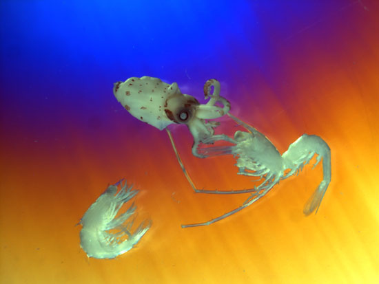
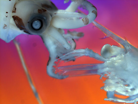
Our good friend Akihiro Shiroza, a marine biologist at NOAA here in Miami, spends most of his working hours looking at the planktonic larvae of fish, corals, crustaceans, etc., under microscope. He was kind enough to share photos of some of the cool plankton he encounters. It appears here that he discovered a squid vs. shrimp battle of micro-mythological proportions. He used a polarizing filter to get the cool rainbow background effect. We’ll look forward to more of Aki’s photographic finds here in the future.

90 Gallon Caribbean Reef Biotope @ University of Miami

Friday, March 21st, 2008

For the past several months we have been working in conjunction with the marine science laboratory at the University of Miami and the UM Aquarium Club (UMAC) in the development of a 90-gallon Caribbean reef biotope in the marine science laboratory. The backstory as to how and why we got involved in this project is more or less as follows.

University of Miami MSC 90-Gallon around it’s heyday (2005-ish)
At that time, a mixed species reef aquarium.

Click to read “Part 1 of 90 Gallon Caribbean reef Biotope @ the University of Miami”…