The flatworms (Convolutriloba retrogemma) featured in the video are shown at 3x normal speed. They each range from 2-4mm in total length.
These particular flatworms harbor symbiotic zooxanthellae in their thin tissue and utilize the excess sugars they create as their primary energy source. Packets of zooxanthellae can be seen as the tiny, red-brown dots along the back of flatworm. Their reliance upon this photosynthesis requires that these flatworms bask in sunlight like little photovoltaic cells, and enables them to live without a developed digestive system.
In the wild, this species can be found in the shallow water of protected lagoons and around mangroves. Reproduction is accomplished asexually via fission, in which the flatworms literally split into two. This strategy enables exponential population growth in optimum conditions. They are the preferred prey of several species of larger flatworms and sea slugs; animals that can tolerate their toxic bodily fluids.
While it appears that the flatworms just glide along like magic carpets, they are actually propelled by invisible cilia (flapping filaments) that slide them across a thin layer of mucous laid over whatever surface they happen to be upon.
Upon close inspection of flatworm-to-flatworm interaction, it is apparent that these flatworms do not like making direct contact with each other. If they do, they react as if stung. It seems that this reaction prevents the worms from piling on top of each other in an effort to gain the best solar power. Instead, they jockey for position until they each find a place in which to ‘park’ themselves, like sunbathers on a crowded beach.
‘The Lynx Nudibranch’ Phidiana lynceus (Lynx Nudibranch) on Spondylus americanus oyster
Music, Video, and Aquarium
2010 Coral Morphologic
Last week we spent a moment making eyes with the oyster (Spondylus americanus). This week we’ll spend a moment with a diverse community of animals and plants that have colonized the upper shell of the very same oyster. Towards the left of the frame is a small colony of flower-like animals known as hydroids. Hydroids are most closely related to jellyfish, but instead remain attached to the reef their whole lives (unlike a jellyfish). But, like the jellyfish, hydroids can pack a powerful stinging punch. The brown, daisy-like creatures seen growing here on the oysters’s back are one such type of hydroid, Myrionema amboinense. This hydroid species derives its brown coloration from the symbiotic zooxanthellae (dinoflagellate ‘algae’) stored in its tissues. The ability to gain nutrition from both prey capture and photosynthesis, allows these hydroids to grow and colonize quickly. The sting from these hydroids is considerably more powerful than that of most corals. The gray, lumpy knobs on the back of the oyster shell are zoanthid polyps, close cousins of the sea anemones. However, these zoanthids are no match against the powerful sting of the hydroids. The zoanthids have all but acknowledged defeat by the encroaching stingers by simply closing up; effectively handing over control of the oyster shell to the hydroids.
‘Oyster Vision’ Spondylus americanus oyster
Music, Video, and Aquarium
2010 Coral Morphologic
Here we look into the face of the thorny oyster (Spondylus americanus). Unlike most shallow-water oyster species, the thorny oyster is a solitary creature that lives permanently cemented to the deeper coral reef. Its fleshy mantle is adorned with sepia-toned psychedelic camouflage that can vary widely from one individual to the next. The rim of the mantle is lined with dozens of eyes that stare out into the depths. These eyes are quite simple, only detecting changes in light that might suggest an incoming predator. If a threat is detected, the oyster will quickly snap its two shells together, sealing the animal inside with its two powerful adductor muscles. It is the adductor muscle that people eat when they eat ‘oysters on the half shell’. Oysters are filter feeders, spending their time siphoning water through gills that strain out particulate matter. As seen in the film, the oyster periodically expels waste and water with a quick contraction of its adductor muscles.
In the second installment (next week) we will explore the upper shell of the oyster and the community of organisms that has colonized it.
‘Transparency’
Unidentified shrimp on unidentified Ricordea polyp
Music, Video, and Aquarium
2010 Coral Morphologic
In the second installment of the ‘Unidentified Ricordea Shrimp’ series we find the (previously featured) unidentified Ricordea shrimp upon an unusual host. While it is is most certainly on a Ricordea polyp, we are not convinced that it is in fact Ricordea florida. Over the years, we have noticed several key morphological and physiological differences that suggest that there are two genetically distinct morphs of Ricordea florida. For practical purposes, we have been referring to the morph of Ricordea seen here (under 470 nm light) as ‘inshore ricordea’.
‘The Arrow Crab’ Stenorhynchus seticornis or ‘Arrow Crab’ guarding a cave entrance
Music, Video, and Aquarium
2010 Coral Morphologic
Take a moment to look into the compound eyes of the arrow crab (Stenorhynchus seticornis). If NASA is looking for a robot capable of navigating rocky planetary terrain, the arrow crab would be a perfect organism to model it after. In the video we look down the sharp, pointed rostrum (‘nose’) of an arrow crab as it appears bobbing in space. In reality, its spindly, spider-like legs are holding it anchored like a sentinel, guarding the opening of a small cave.
Arrow crabs are an abundant species on Floridian reefs, living perched near cracks and crevices in coral heads where they can retreat if threatened. Their pointed rostrum, triangular body, and protruding eyes gives this crab the appearance of a predatory lizard fish that can dash away at a moment’s notice. Instead, the arrow crab is rather slow moving, relying on the fact that the paucity of meat inside the spiny, twig-like exoskeleton of the arrow crab makes it unappetizing to a would-be-predator. This unique anatomical configuration likely explains their abundance in the wild.
Like other decapod crustaceans, the arrow crab has 10 legs (8 walking legs, and 2 pincers or ‘chelipeds’ properly). However, if you look carefully, you’ll notice that this particular crab is missing the last leg on the right side of its body. Fortunately, crustaceans are capable of regrowing amputated legs. Only a few hours after it was filmed, this arrow crab molted, and as if by magic, regenerated its tenth limb.
‘The Fire Coral’ Pt. 1
A feeding Balanus sp. barnacle encrusted by Millepora alcicornis
Music, Video, and Aquarium
2010 Coral Morphologic
Millepora alcicornis, or fire coral, is not actually a true coral, but a hydrocoral. Hydrocorals are colonies of hydroids that secrete a shared limestone skeleton, making them more closely related to jellyfish than true corals. Here in Florida, fire coral is extremely abundant on our reefs where they serve as the underwater equivalent of a sunburn to unsuspecting divers. Skin contact with fire coral will result in immediate burning pain, followed by an itchy welt that can last for several days.
Fire coral is frequently found encrusting over neighboring corals, starting from the bottom and slowly killing the coral until the colony is completely encased in living limestone. Because fire coral contains symbiotic zooxanthellae (like most tropical stony corals), they are capable of fast growth rates that help build a coral reef. Upon close inspection of fire coral, the stinging polyps can be seen as needle-like projections. At even closer magnification, grape-like bunches of stinging nematocysts can been seen protruding along the polyps’ length. These polyps are retractable, and when an edible food particle is captured, it can be drawn back towards one of the many mouths that dot the surface of the colony. In the video we see a colony of barnacle shells (Balanus sp.) that have been encrusted by fire coral. Unlike the corals though, the barnacle can continue to live beneath the veneer of fire coral.
Barnacles are most commonly found living in the inter-tidal zone where they live periodic lifestyles of low tide rest and high tide activity. When immersed in water, the barnacle feeds with legs specialized for feeding called cirri. The cirri are covered with comb-like filaments that rake the water for passing plankton. If a particle is caught in the cirri, it is drawn back to the animal’s mouth and eaten. When barnacle larvae settle out of the floating plankton themselves, they permanently affix themselves to a life-long location. Barnacles have a special ‘cement gland’ under their bodies that produces an impressive proteinaceous adhesive that holds the animal firmly, in spite of the heaviest of waves. A series of calcareous plates (commonly six) form a turret that protects their soft bodily tissues from predators. Despite their simple appearance, barnacles are in fact crustaceans, like shrimp, crabs, and lobsters.
‘The Sun Coral’
The feeding of a Tubastrea coccinea coral cluster
Music, Video, and Aquarium
2010 Coral Morphologic
This week’s video features a colony of Tubastrea coccinea coral polyp clones feeding on passing zooplankton. The film is sped up 10 times to emphasize the feeding abilities and coordination between the sticky tentacles and the polyps’ mouths.
Tubastrea coccinea or ‘Sun Corals’, have an unusual background story, being the only invasive stony coral to become established in the Caribbean basin. Native to the tropical Indo-Pacific Oceans, they were first noted living on ships’ hulls in Puerto Rico and Curacao (Southern Caribbean) in the mid 1940’s. Over the ensuing decades, they eventually spread elsewhere throughout the entire Caribbean and Gulf of Mexico on the prevailing water currents. It is believed that these sun corals may have originally entered our region as larval stow-aways in the ballast water of intercontinental ships that passed through the Panama Canal.
‘Lima scabra’
The tentacles and mantle of a Lima scabra file clam filter feeding
Music, Video, and Aquarium
2010 Coral Morphologic
Lima scabra is a common resident on Floridian and Caribbean reefs where it can be found wedged in crevices, with only its long tentacles extending out into the water column. Usually these tentacles are crimson red (as seen in the specimen above), although they are occasionally white in color. Lima scabra can grow to about 3.5 (9cm) inches long.
Like most bivalves, Lima scabra is a filter feeder. It siphons water in through its fleshy mantle (seen in the video), and strains any edible particulate matter before pumping the water back it out. It holds itself semi-permanently in place through the use of ‘byssus threads’. The threads are formed by a viscous protein secretion that cures instantly upon contact with seawater. These byssus threads have captured the attention of bio-engineers who seek to replicate their strong adhesive properties for industrial applications. However, if the clam comes under attack from a predator, it is capable of detaching and swimming away. They can move surprisingly quick; swimming in fast, jerky movements, propelled by the repeated snapping-together of its shell.
‘Purple Forest’
Decorator Crab (Microphrys bicornuta) on Asparagopsis taxiformis algae
Music, Video, and Aquarium
2010 Coral Morphologic
This week’s video features an aquascape comprised of the beautiful purple macro algae Asparagopsis taxiformis. However, if you pay close attention to the left 1/3 of the screen, you’ll notice something… moving with claws. Nestled amongst the algae is a perfectly camouflaged decorator crab (Microphrys bicornuta). Keep paying attention… at 26 seconds into the clip you’ll notice a tiny isopod crustacean float by in the current and descend helicopter-style right onto the crab’s back. The unsuspecting isopod has no idea that it has landed upon an algae covered beast. Furthermore, it appears that the crab is not aware of the unexpected visitor until the isopod begins to explore its decorated exoskeleton. 50 seconds into the clip the isopod meets its fate with a few swift snatches of the crab’s claws. Without missing a beat, the crab continues scavenging amongst the rocks and algae. And life on the reef goes on.
Decorator crabs are amazing creatures in that they pick up pieces of their surrounding habitat and place them on their carapace (back, exoskeleton) in order to blend into their surroundings. Decorator crabs that live amongst sponges decorate with sponges, those that live amongst zoanthids use zoanthids, and so on. This instinctual logic is truly remarkable. The crab in the video has attached small pieces of the Asparagopsis upon itself, and as a result is all but indistinguishable from its surroundings.
‘Corynactis viridis’
The feeding of a Corynactis viridis corallimorph
Music, Video, and Aquarium
2010 Coral Morphologic
This past week we finally received our long awaited Corynactis viridis from our good friend Dr. Yvan Perez at the Institut Méditerranéen d’Ecologie in Marseilles, France. I collected these polyps this past June while diving in the Mediterranean, and Dr. Perez has been culturing them in his lab in the interim. Laurent Foure, the current curator of the Noumea Aquarium in New Caledonia, also collected us several stunning morphs from a different Mediterranean location before he left for the South Pacific.
Corynactis viridis is an archetypal corallimorph species found all along the cold, rocky coastlines of Western Europe. Its distribution across the Mediterranean is much more sporadic and considerably less common. They are frequently referred to as ‘jewel anemones’, which is a misnomer, as they are not anemones despite their superficial resemblance. Typically, polyps range in size from about 3-10mm in diameter and can be found in a seemingly limitless number of color morphs. What they lack in individual size, however, they make up for in colonial dominance. It is not uncommon for colonies to completely carpet large areas, frequently rocky outcrops and vertical surfaces. As these colonies are comprised of clones, this carpet will be of uniform coloration, creating the illusion of a singular connected organism. Multiple color morphs will often be found living in close proximity, creating a technicolor patchwork of tiny individuals. Their colors are often vibrant with fluorescent accents. Unlike most of our tropical corallimorphs, C. viridis are non-photosynthetic, relying entirely on the capture of plankton by their sticky tentacles. At the end of each tentacle is a small ball known as an acrosphere; a tell-tale characteristic of all non-photosynthetic corallimorphs.
In this video a single Corynactis viridis polyp (about 8mm in diameter) is seen capturing and digesting tiny plankton as they flow past in the current. As the tentacles capture food, they retract towards the animal’s mouth, located at the center of the polyp. The mouth is likewise transformable; capable of extending, expanding, and enveloping food items. The total elapsed time was roughly 12 minutes and sped up 1200% in order to demonstrate the hydraulic muscular contractions and contortions that the polyp goes through while feeding. 470nm LED light is used to highlight the fluorescent orange ring around the outer diameter of the polyp.
‘The Christmas Tree Worm’
Spirobranchus giganteus – Amber Morph
Music, Video, and Aquarium
2010 Coral Morphologic
Christmas tree worms (Spirobranchus giganteus) are an abundant creature on Floridian reefs, making their permanent homes encased inside the limestone skeletons of live coral. Found in a seemingly endless variety of colors and measuring 2-3 cm in diameter, dozens of these worms will typically adorn massive coral heads in local waters.
Using only the perception of light and vibration, these animals will retract at lighting speed at the first sense of something ominous approaching. Fortunately the worms come equipped with a a protective double-horned operculum that seals the worm safely inside the impenetrable coral. A sharp, calcareous spike extends forward of the tube’s opening, acting as a further deterrent to a would-be predator.
The spiraled, ‘branchial crown’ serves as both breathing and feeding apparatus for the worm, and is the only part of the worm’s body that is extended into the water column. The feathery appendages, known radioles, collect plankton that drift by in the current. The radioles are lined with cilia that direct the captured food down the spiral to the worm’s mouth.
‘Preener’
Mithraculus cinctimanus crab on Ricordea florida corallimorphs
Music, Video, and Aquarium
2010 Coral Morphologic
Shown above is a 1cm Mithraculus cinctimanus, commonly known as the banded clinging crab. Typically this species is known to live in association with a variety of Caribbean sea anemone species. However, several years ago we noticed that juvenile and sub-adult banded clinging crabs seemed to prefer the protection amongst Ricordea florida polyps in the wild. When they are small, like this one, the carapace (shell) of the crab is nearly entirely covered by a fuzzy red algal camouflage. As they get larger (up to 25mm) they lose much of this hairy coat, revealing a striking white and maroon patterned exoskeleton.
The video shows the crab alternating between preening its own algae covered carapace and the fluorescent tentacles of the Ricordea florida on which it lives. It is possible that the crab may ingest some of the polyps’ mucus as an occasional food source. The video was sped up considerably (9x). At normal speed the polyps appear static, but at this speed the regular hydraulic undulations and contractions of the R. florida polyps are clearly visible.
‘Unidentified Ricordea Shrimp’
Unidentified commensal shrimp on Ricordea florida corallimorphs
Music, Video, and Aquarium
2010 Coral Morphologic
Shown above is the first documented video of a currently unidentified shrimp commensal with Ricordea florida corallimorphs. The nearly invisible shrimp measures only 9mm in total length. The ricordea polyp is about 30mm in diameter for comparison. We first reported and photographed this shrimp in October 2009. Subsequently, we sent a preserved specimen to taxonomist Dr. Richard Heard at the Gulf Coast Research Laboratory. Dr. Heard was unable to match the specimen with a described shrimp species. We will be sending additional specimens in the near future to confirm this shrimp’s newness to science.
‘Cleaner Pt. 2’ Periclemenes pedersoni shrimp on Ricordea florida corallimorphs
Music, Video, and Aquarium
2009 Coral Morphologic
Here a 2 centimeter Periclemenes pedersoni shrimp is seen perched on its host colony of fluorescent orange Ricordea florida corallimorphs. Pederson’s shrimp are native to Floridian and Caribbean waters, and are found in symbiotic association with a variety of sea anemone and corallimorph species. Pederson’s shrimp live amongst the tentacles of these animals, immune from their sting. They act as ‘cleaners’ of much larger fish. The shrimp wave their antennae and swim in a characteristic swaying motion to signal that they are ‘open for business’. Fish seem to learn and remember the specific location of these ‘cleaning stations’, returning as needed for service. When not removing parasites from fish, these shrimp will forage for bits of food amongst its host’s tentacles and spend time preening itself, as can be observed in the video. Pederson’s shrimp form monogamous mated pairs that will live together on same host. These shrimp are quite territorial, fighting fiercely with its own kind if another Pederson’s shrimp tries to take up residence on its anemone or corallimorph. If an ‘unmated’ shrimp tries to take up residence too close to an established territory, the invader will usually be dissuaded through threat displays and physical harassment.
Pederson’s shrimp are closely related to the spotted cleaner shrimp (Periclimenes yucatanicus) featured last month.
During the nights of Art Basel Miami Beach our 2009 film reel was displayed on the 80′ X 42′ LED screen on the facade of the American Airlines Arena in downtown Miami as part of the Media Mesh Project. The video compilation was curated by artist George Sanchez-Calderon and featured short films by the following artists: Lou Ann Colodny, Aiden Dillard, Alvaro Ilizarbe (Freegums), Justin Long, Ferran Martin, Coral Morphologic, Gavin Perry, Carlos Rigau, Bert Rodriguez, Damian Rojo, TM Sisters, and Jen Stark.
J.D. McKay (left) and Colin Foord (right) of Coral Morphologic; a self portrait.
During the night of Saturday, December 5th we displayed a video projection at the Art Basel Miami Beach satellite Moksha Art Fair. The subject of the the film was an Entacmea quadricolor sea anemone (right) and its corresponding reflection (left).
Pictured above and below is ‘Version Key’ as installed at the Miami’s Independent Thinkers art show during the days of Art Basel Miami Beach. The aquarium consisted of an eroded cinder block, red mangrove (Rhizophora mangle) seedling, and three upside-down jellyfish (Cassiopea xamachana) pulsating on oolitic sand; not an atypical South Floridian assemblage. It was displayed against a silver parachute backdrop on a concrete block pedestal surrounded by oolitic sand.
Cuddle Fish is the creation of Miami contemporary artist Bhakti Baxter. The limited-edition zine, also compiled and printed by Baxter, features images contributed by a collection of his peers. The cover image is a Sepia sp. Cuttlefish that Colin photographed back in 2007 in Tulamben, Bali. You can find this zine in physical form at happenings around Miami during Art Basel Miami Beach 2009.
CORAL MORPHOLOGIC Art Basel Miami Beach 2009 schedule:
‘Cleaner Pt. 1’
Periclimenes yucatanicus shrimp on Condylactis gigantea sea anemone
Music, Video, and Aquarium
2009 Coral Morphologic
Here a three centimeter Periclimenes yucatanicus (spotted cleaner shrimp) preens its exoskeleton and gills amongst the tentacles of a Condylactis gigantea sea anemone. P. yucatanicus is a relatively common species that lives in association with sea anemones and corallimorphs here in Florida and throughout the Caribbean. The stinging tentacles of their hosts provide them with protection from would-be predators. However, despite its vulnerable size, these shrimp act as ‘cleaners’ of larger fish. They remove and consume any parasites and dead tissue they find, often from the fish’s teeth. These fish clearly perceive the benefits of the shrimp’s cleaning abilities over its value as a tiny morsel. This particular Condylactis gigantea has a fluorescent green oral disc which is unusual for the species.
Recently while diving off of Key West, I was fortunate to come upon a rare and unidentified species of Palythoa. This was the first time that I have come upon this type in five years of frequent diving throughout the Florida Keys. Apparently it is less rare elsewhere in the Caribbean, but as of now has yet to be properly identified by a zoanthid taxonomist. On its particular patch of reef it was relatively abundant, despite being completely absent in seemingly identical reefs in the surrounding area. And while it might seem logical that it only takes one lucky zoanthid larvae to ultimately colonize a large area, it seems that there were at least 2 separate morphs cohabiting the area, which makes its complete absence from other nearby reefs compelling. And while Caribbean Palythoa display morphologies that seem to overlap, these particular Palythoa have a few traits that make it noticeably distinct:
Small size (1/4″-3/8″ disc diameter)
Translucent oral disc, often with teal-bluish iridescent sheen
Distinctive white splotch, often butterfly shaped
Eyelash-like tentacles
But considering that the morphologies of Caribbean Palythoa species seem to blur together, genetic analysis will be the most reliable way to determine species-hood. Fortunately our friend James Reimer is a zoanthid expert in Japan’s Ryuku Islands with access to such equipment and expertise. In addition to sending him samples of this Palythoa morph, we will also include a variety of other local Palythoa morphs to see just how distinct the individual species are.
Pictured above is a very tiny (10mm) shrimp that lives commensally with Ricordea florida polyps.
Over the past several years I have occasionally encountered fleeting glimpses of tiny shrimp that live amongst the pseudo-tentacles of Ricordea florida. On all the previous occasions that encountered one, I had never been properly equipped with a super-macro camera kit. A dive this past September finally warranted a good photo. Ricordea shrimp are tiny (8-12mm) and nearly transparent, making them very difficult to detect. It is unlikely that these might be juveniles of a more common commensal species (e.g. Periclimenes pedersoni, P. rathbunae, or P. yucatanicus), as it is clear from the photo above (and from recently collected specimens) that they are mature egg-bearing females at this small size. If you look closely at the photo, you’ll notice in the upper left-hand side of the photo that there is another pair of eyes. At the time of the photo I didn’t notice that there were several other tiny and completely clear shrimp living with this female. It was only while viewing the photos close-up that I noticed these other shrimp. Most likely they are male or juvenile females living colonially. On subsequent dive trips I have found several groups (3-5) of these tiny shrimp all living on the same R. florida colony.
Today we are sending off a preserved specimen to Periclimines shrimp expert Dr. Stephen Spotte for taxonomic inspection. He will be able to determine whether this shrimp has been previously identified, or whether we are dealing with a new species altogether.
An unusual colony of Discosoma sanctithomae with perfectly spherical vesicles from the Florida Keys at 10m of depth. Note the turbid sea floor, characteristic of this species’preferred habitat.
The unusual spherical vesicles of these Discosoma sanctithomae polyps once gave this morph a separate species designation Orinia torpida by Duchassaing & Michelotti in 1860. Despite several other taxonomists re-examining the single preserved specimen in the Zoological Museum of Turin in the first half of the 20th century, it was the late great corallimorph taxonomist J. C. Den Hartog who finally corrected this error in 1980. It is understandable that such confusion could occur. Until the advent of scuba diving, many taxonomists would never actually observe the living marine animals they classified. Instead these Euro-centric scientists would rely on preserved specimens sent back to them from various collection missions around the world. With few specimen samples to compare with, morphologic oddities like the pictured polyps could easily be considered to be something entirely new.
A close-up of a staghorn coral (Acropora cervicornis) branch tip. Note the healthy coral polyp extension.
This past Saturday I took my friend Jeremy (of coralpedia.org) and his wife on a shore dive off Fort Lauderdale Beach. I had heard rumors that decent stands of the endangered staghorn coral (Acropora cervicornis) were abundant in this area. Despite (relatively) poor visibility, we found these rumors to be correct. Acropora cervicornis was one of, if not the most, common stony corals only 250-350 meters offshore this popular sunbathing mecca in only 6-7 meters of water. It is amazing to see how even a small ‘bush’ of A. cervicornis can attract dozens of small fish seeking refuge amongst its branches. It is clear that the widespread die-off of this single species has had a detrimental impact on the entire ecosystem of the Florida’s coral reefs. I can only imagine what the reefs in the Florida Keys were like 30 years ago, as today most are just lumpy humps of rock dominated by massive coral heads, gorgonians, and macroalgae. The interstices of A. cervicornis branches provides a habitat that is unmatched. Seeing an abundance of A. cervicornis so close to shore in Fort Lauderdale is encouraging.
The spongy decorator crab (Marcocoeloma trispinosum) takes expert care in snipping off pieces of living sponge and attaching them to its carapace (exoskeleton shell). Detritus and debris are added for additional camouflaging effect. This particular crab has taken the decoration to the next level by including some spectacular zoanthids (Zoanthus sp.) into its design. As you can imagine, the crab was living amongst a colony of the same zoanthid morph, rendering it nearly impossible to detect. Furthermore, decorator crabs move very slowly and deliberately, quite unlike the unpredictable scurry of most other crabs. The purposeful addition of camouflaging marine life to the body of this crab highlights the evolution and subconscious intelligence of ‘tool-use’ at such a ‘primitive’ level in the animal kingdom.
Still photo (above) of aquarium/ projection of our contribution to the Sweat Records X Iggy Pop t-shirt release party, August 22, 2009 at the Awarehouse in the Wynwood Art District. This installation consists of a 10″ X 10″ X 4″ acrylic aquarium atop an overhead projector projecting a 15′ X 15′ image onto the courtyard wall. The live-action component of the installation features a performance by thirty photosynthetic jellyfish (Cassiopea xamachana) collected in the mangrove estuary near the Virginia Key Wastewater Treatment Plant, here in Miami, FL.
A close up of a colony of Pectinatella magnifica zooids; a freshwater bryozoan.
Pectinatella magnifica is another example of a coral reef cousin that I found living in a freshwater lake in Maine, alongside the previously featured Spongilla sp. sponge. From a distance, the colonies that these animals create appear as large jelly-like masses that might be confused with frog’s eggs (see photo below). They are typically found encrusting submerged tree branches, plants, and rocks. Most bryozoans are found in saltwater, but a few species such as P. magnifica are found in freshwater habitats. It would be forgivable to call the individual animals ‘polyps’, as they are superficially coral-like. However, unlike cnidarian (coral-like animals) polyps, bryozoan zooids have a complete digestive system with a separate mouth and anus, whereas cnidarian polyps have a single mouth/ anus opening.
If you look closely at the macro photo you will notice a couple of worms that are living in casings between the valleys that separate sub-colonial groupings of the zooids. The worms were wriggling constantly, most likely in an effort to create a water flow through their tubes that would draw in food items. It would be interesting to know whether these worms are specific only to Pectinatella magnifica, or whether they are simply opportunistic.
The full-sized colony of Pectinatella magnifica looks and feels a lot like a mass of frog’s eggs. It has encrusted over a submerged pine twig.
This freshwater sponge (Spongilla sp.) was found encrusting a submerged tree branch near the shoreline of a small lake in Maine. Note the small oval arthropods living on the surface of the sponge near the middle of the photo.
I recently returned from a short vacation to New Hampshire/ Maine to see family and stomp the formative grounds (and waters) of my youth. While paddling around the small lake that I spent my summers, I found an abundance of this freshwater sponge (Spongilla sp.) growing submerged near the shoreline. Growing on rocks it has a flat, encrusting morphology. On submerged branches it forms lumpy encrustations. And on shallow muddy bottoms it sends up skinny tendrils. The tissue is colored bright green due to a symbiotic association with algae. While you might find yourself thinking “freshwater sponge? really?”, these sponges can be found across most of North America from Alaska south to Florida in relatively still freshwater lakes and ponds. As a testament to their adaptability, this sponge can ‘degenerate’ into a dormant state, forming tiny cysts called gemmules. These gemmules act as ‘seeds’ when favorable conditions return. The sponges reproduce sexually in the summertime and release free swimming larvae. Next time you find yourself at a lake, take a closer look at what you might otherwise think is just some lumpy ‘algae’.
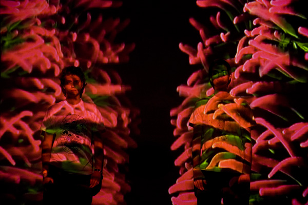
Still photo (above) of aquarium/ projection of our contribution to The Collabo Show, July 25, 2009; an installation dubbed ‘Cassiopeia 1’, featuring photosynthetic jellyfish (Cassiopea xamachana) collected in the mangrove estuary near the Virginia Key Wastewater Treatment Plant, here in Miami, FL.
Saturday, July 25th we will be exhibiting a live aquarium and video projection in The Collabo Show, a one-night only showcase of contemporary Miami artists. 85 NW 71 Street, Little Haiti. 7PM – 12AM.


")
")