coral morphologic header


Author Archive

Fluorescent Pycnogonid (Sea Spider)

Tuesday, April 7th, 2009

Morphologic Studios 2009 Colin Foord

The cephalothorax of this unidentified pycnogonid is covered in wisps of  cyanobacteria (perhaps as camouflage) which itself fluoresces orange-red. This specimen measures about 1 cm in diameter. Fluorescence photograph.

It is amazing the wide variety of sea creatures that demonstrate fluorescence. Animals that you would never suspect to “glow” do just that when illuminated with the proper wavelength light. For the past several years I have been using an underwater flashlight that I custom outfitted with 470nm blue LEDs in order to be able to scan for glowing creatures in the darkness. This pycnogonid sea spider (not an arachnid) is a prime example of an unexpected life form displaying an ability to fluoresce. The “spider web”-like pattern of fluorescence over this specimen is truly impressive.

Pycnogonids, commonly known as “sea spiders” are rarely seen in the wild (but not necessarily rare), and are an unusual class of arthropod. Most are very small (<1cm), and hence overlooked. However, in the Antarctic there are some monsters that can grow up to 90cm (Pycnogonaphobia?)! One of their most distinctive features is that they possess a proboscis (a drinking straw if you will) that allows them to suck out fluids from soft-bodied invertebrates such as corals, anemones, and corallimorphs. In the wild they rarely do lasting damage to their prey. However within the confines of an aquarium I could imagine that this might not be the case where their selection of hosts is limited. As such, they should be regarded as potential parasites of corals within the aquarium, and removed if detected. Despite this threat, they don’t seem to be widespread pests within the aquarium keeping world.

Morphologic Studios 2009 Colin Foord

The same pycnogonid specimen in a defensive posture after being prodded “into position” for photography.

A Walk Through Big Cypress

Wednesday, January 14th, 2009

If you ever find yourself having to cross the southern portion of Florida, you’ll have the privelege of transecting the Florida Everglades. For most people, this involves taking I-75, aka “Alligator Alley”, which is four lanes wide and moves quickly when there isn’t a major hold-up (which is somehow more often that you’d like). Given that the flow of traffic usually moves in excess of 80 mph, it is quite unlikely that you will get to see any of its namesake reptiles.

A much more scenic route, that will actually reward you with an alligator or four, is to take the Tamiami Trail, aka Highway 41. The road is only two lanes, but still manages to cruise at a comfortable 60 mph with relatively few hold ups or traffic. The Tamiami Trail crosses Florida to the south of I-75, and takes you through a picturesque cross-section of the Everglades. Along the way you’ll find rest stops, nature trails, and Miccosukee-owned road side attractions that give this route great Floridian character.

(more…)

Miccosukee Swampwalk

Wednesday, October 22nd, 2008

I spent this past Sunday morning slogging through the eastern edge of the Miccosukee Everglades. The Everglades, at the tail end of a bountiful rainy season, are finally looking like the river of grass that they ought to be. For the past several years South Florida has been in a serious drought, but this year has brought us a much needed replenishment of rainwater.

More photos below the jump…

(more…)

Rest in Peace(s)… ‘Discosoma 1.0’

Sunday, September 21st, 2008

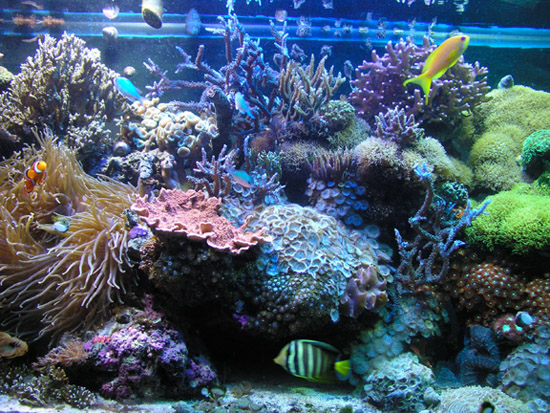
We were disheartened to receive a call from our good friend Lauren Reskin, owner of Sweat Records, this past Sunday with the news that her store had been broken into, thieved, and trashed. The worst karmic act committed was the wanton destruction of the Red Sea Max that we installed and maintained as a gift to her store and our friends that frequented it. We set up the aquarium at the very end of November 2007 for the opening of Sweat’s third incarnation here in Miami.

According to the police, the thieves cut through the security gates of the store’s back entrance early Sunday morning and proceeded to steal the store’s computer, electronic equipment, several pieces of art from the walls and… knock over the aquarium.

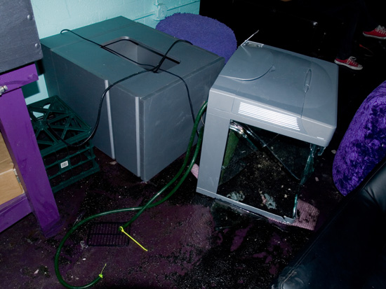
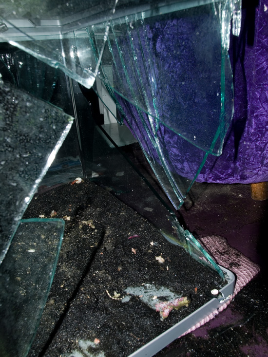
This what we arrived to find (minus the live rock and corals that we had already gathered up by this point in our effort to revive what we could.

Sweat Records was closed on Sunday, so it wasn’t until almost 9 pm Sunday night that Lauren happened to be driving by her store and noticed the break in. It is likely that the contents of the aquarium were dry for over 18 hours.

We arrived at Sweat Records in a daze, unable to believe that someone would go through the trouble of destroying an aquarium just for the thrill of instant destruction, until we saw it with our own eyes. We were expecting the worst; that nothing could possibly still be alive.

All of the fish were dead. We hypothesize that the impact and subsequent shock-wave of the aquarium shattering against the floor must have killed them immediately as there was several inches of standing-water still on the floor that could have otherwise provided them with refuge.

However, there was some good news. Both of the harlequin serpent stars and a porcelain crab showed minimal signs of life, despite being nearly dry. When plopped into a plastic bag with water, they revived within a few minutes. The snails and blue leg hermit crabs all survived. We were initially highly skeptical that the Discosoma sp. corallimorphs would survive 18+ hours out of water, but we gathered up all of the pieces of live rock and brought them back to our facility anyhow. We didn’t want to put them into our holding or grow-out systems, as we were afraid that if they died as we expected, they would only foul the water. Instead, we placed them in two 18 gallon mortar tubs with heavy aeration and a power head and hoped for the best.

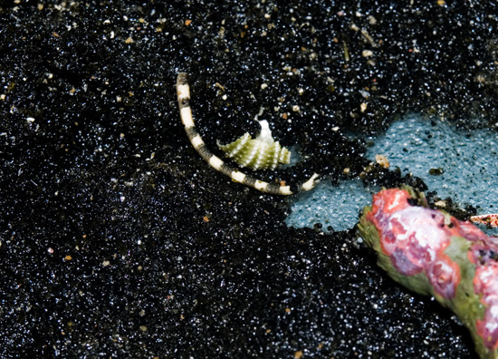
The severed arm of a harlequin serpent star is seen above. Fortunately, the serpent survived the ordeal and more than 18 hours out of water. It will regenerate this arm in short time. The fish weren’t so lucky.

To attest to the ruggedness of the Red Sea Max, the hood and even the light bulbs remained undamaged despite the brute force impact. Likewise, the RSM’s silver stand escaped even minor scratches despite landing face down. The two return pumps were the only other parts of the RSM beside the actual glass aquarium that had to be thrown out. Because the electrical cords never pulled out of the wall, they continued to run dry after the fall.  This caused them to overheat and melt together! The Current 1/10hp chiller managed to unplug itself and therefore survived with nary a blemish.

We were relieved the following morning to find that most of the corallimorphs were recovering nicely. Now, almost a week later, they have nearly made a complete recovery. We did lose a few polyps, but I’d estimate at least 80% survivorship.

Unfortunately, we did lose about half of the pink coralline algae from the live rock. Coralline algae does the most amazing thing when it dies; it fluoresces a bubble gum pink color. While temporarily beautiful, it soon turns to a dead white bleached state within a day or so. If anyone knows why this phenomenon occurs, please inform us. To lose this much coralline algae cover from your live rock after nearly a year of cultivation is a real bummer.

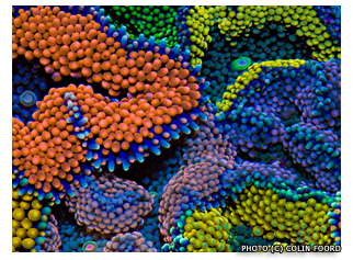
You can see a fluorescence photograph of the live rock below:

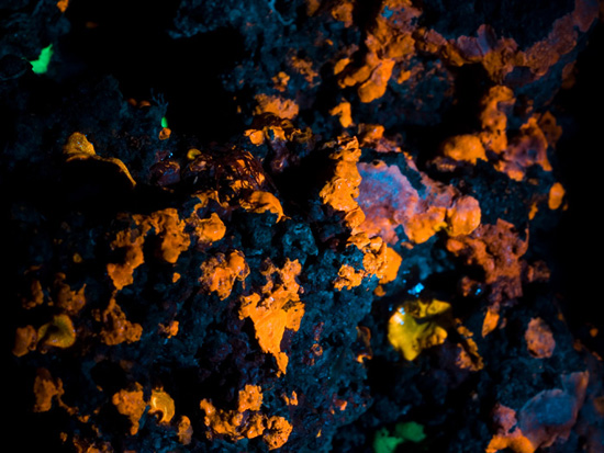
The dying pink crustose coralline algae fluoresces after death. The photograph was taken under blue wavelength (450 nm) light to maximize fluorescence. Several gold and green Discosoma sp. can also be seen fluorescing, despite 18 hours out of water.

While dismayed at losing this aquarium, and just as it was maturing into an exceptional little reef aquarium, we are not deterred. Sweat Records will be holding a series of benefit concerts in the next several weeks to help raise money for a better security system and a new aquarium. We hope to have another aquarium up and running by the time of Sweat’s first anniversary of being in their current location. We’ll keep you posted.

Here are a couple of articles written by local blogs about the break in:

Miami New Times

ARTLURKER

A Freshwater State of Mind

Thursday, August 14th, 2008

Pictured above is an aquarium that Jared and I established in late 2004. It is currently maintained most capably by Jared’s father Dennison in New Hampshire. It is stocked entirely with plants that originated from a 150 gallon angelfish (Pterophyllum scalare) aquarium I had enjoyed in Miami from 2003-2004. The dominant plants are Cryptocoryne wendtii, Crinum thaianum, and Microsorum pteropus (Java fern). All of these plants are hardy, and don’t require much special care. The aquarium is stocked with a variety of standard “community” freshwater fish such as tetras, platys, and rasboras. This aquarium is a nice example of a natural, planted aquarium that requires a low degree of maintenance, yet rewards with a high amount of peaceful satisfaction.

Neptune Memorial Reef

Friday, August 1st, 2008

Today my good friend Carlos and I made a morning dive excursion off of Miami to visit the Neptune Memorial Reef. I had heard about this artificial reef quite a while ago when it had been originally proposed to simply be a “replica” of Atlantis. It seems that the goals of the project shifted towards a more realistic and profitable goal by becoming the world’s first underwater cemetery and memorial park. The first phase of construction was completed in November 2007, and I’ve been looking forward to taking a first-hand look ever since.

(more…)

Incredible Image

Monday, July 21st, 2008

I picked up a lot of free promotional material at the ICRS (my personal favorite being a 6 foot National Geographic world map detailing where all the worlds coral reefs are). This little post card advertising the 2nd Asia-Pacific Coral Reef Symposium really caught my eye. A perfectly-balanced tabular Acropora with a staghorn Acropora species growing in the center. Most excellent.

Coral Mucus and Bacteria: A Symbiosis?

Monday, July 14th, 2008

Anyone who has ever scraped or cut their skin on a living coral can attest to the malignant nature of what should otherwise be a minor abrasion or cut. These scrapes don’t heal very quickly, and can become infected very easily. The reason is that the mucus coating secreted by the coral harbors a dense population of bacteria that apparently gains protection and nutrition from said mucous. The relationship between mucous bacteria and coral is only now beginning to be unraveled.

That there were at least 16 presentations and posters that focused specifically on coral mucus at the ICRS, shows the level of interest this topic has been receiving within the marine biological world.

Several of the research projects concluded that the bacterial populations within the coral mucus are in fact mostly unique and independent of the bacterial populations that are found in nearby environments (surface sediments, biofilms, water column, etc). This indicates that the coral mucus/ bacteria relationship is more complex and specific than previously thought.

An important research topic on coral mucus bacteria revolves around their relationship to coral immune health and disease prevention. It is speculated that the mucus bacteria are somehow capable of thwarting coral infections (other bacteria, protozoans, etc), by maintaining a balanced population within the mucus (perhaps a similar theory of using ‘probiotics’ as preventative measure?). Whatever the mechanism, it appears that the coral mucus and resident bacteria population acts as a protective barrier against pathogenic invaders.

Another important role of coral mucus is to act as a medium for nutrient transport. I assume that this perhaps relates to the mucus’ ability to help adhere to and coat food particles, thereby aiding and enabling digestion. This is a conjecture on my part, but it seems like a logical process.

A research poster entitled “A Quantitative Approach Linking Coral Mucus And Their Symbiotic Zooxanthellae in Response To Environmental Change” found that 45% of the daily fixed carbon (i.e. the food produced from photosynthesis), was incorporated into coral mucus in Montastrea annularis. This demonstrates the vital importance that coral polyps place on mucus production.

In the poster mentioned above, the researchers determined that as water temperatures were increased by 1.5 degrees C, mucocyte density (specialized cells that produce mucus) increased, while zooxanthellae density decreased. They draw the conclusion that increasing temperatures cause M. annularis to rely more upon heterotrophy (eating), than upon autotrophy (zooxanthellate photosynthesis). Bleached corals were found to have lower densities of mucocytes, but the remaining mucocytes were greatly enlarged, indicative of highly increased mucus production per mucocyte.

A thought that popped into my head while reading the results of this paper, in combination with the other information I picked up in several lectures on the topic, is that perhaps the coral actually digests the bacteria that live in the mucus layer, thereby adding an additional symbiotic food source (Zooxanthellae being the other “food” producer). It seems possible that by providing a suitable medium for bacterial growth, the coral is able to culture its own “bacteria garden” that is consumed at a rate that is balanced with mucus production and the bacterial growth within it.

I hope that future research continues to look into this area, as it is possible that there is still a piece of the coral nutrition puzzle that is still waiting to be unraveled.

Corallimorpharians, etc. @ ICRS

Friday, July 11th, 2008

There has been recent interest in the corallimorpharians by marine biologists, due to genetic studies that demonstrate that some hard corals (Scleractinia) are actually more closely related to corallimorphs than to other members of Scleractinia. This has resulted in several papers that suggest that corallimorphs once had a Scleractinian ancestor with a calcium carbonate skeleton. Prior to these genetic studies, it had been suggested (through taxonomic studies) that corallimorphs were in fact more closely related to sea anemones (Actiniaria).

To explain why the loss of this skeleton may have occurred, researchers have been suggesting that increased levels of carbon dioxide during the Triassic period (from about 200-240 million years ago), would have caused a reduction in oceanic pH, which resulted in a decreased ability by hard corals to build a calcium carbonate skeleton. These conditions would therefore select for corals that could adapt to live without their skeletons. And voila!, the corallimorpharians were born. Unfortunately, the fossil record of corallimorpharians is minimal due to their lack of calcium carbonate skeleton, making this claim hard to prove. It is certainly a very seductive argument, especially now that atmospheric levels of CO2 are once again rising to levels that may soon have a detrimental impact on hard corals and their ability to continue reef building. This has some scientists positing that in the future as hard corals start dying off, soft bodied anthozoans will once again have an adaptive advantage, resulting in their proliferation.

An interesting poster entitled “Mechanisms of Microhabitat Segregation among Corallimorpharians”, examined the ability of two common Red Sea corallimorphs to handle UV light stress. One species Rhodactis rhodostoma, is commonly found in shallow-water reef flats where light intensity is very high. The other, Discosoma unguja, is common at deeper depths in shaded areas. The researchers exposed both species to identical conditions of light with several variable treatments (high light, shaded, etc.).

To understand what the researchers were looking for, as far as “light stress” is concerned, one must understand what happens within the corals’ tissues and zooxanthellae during photosynthesis. To summarize, one byproduct of zooxanthellae photosynthesis is the formation of highly reactive and potentially damaging oxygen ions. The coral must deactivate these harmful byproducts in order to prevent tissue damage. Fortunately, corals have evolved certain enzymes that are capable of deactivating these oxygen radicals. This is the reason why it is very easy to “light shock” your corals by rapidly increasing the amount of light they receive. The coral is simply unprepared to deactivate all of the additional harmful byproducts of the increased photosynthetic activity.   typical emergency reaction by the coral is to expel its zooxanthellae  (bleaching) so that no further damage can occur.

Anyway, back to the study at hand. When the Discosoma unguja were placed under full strength light, they showed a decided inability to handle the affects of increased photosynthetic activity. Instead of an enzymatic ability to deal with high light, they expressed an ability to move away from the light source. Crafty little creatures.

Rhodactis rhodostoma, on the other hand, proved to be resilient to high levels of light. Tissue analysis showed much higher levels of the enzymes necessary for deactivating harmful oxygen ions than in D. unguja.

What the study did not mention however, was whether the Discosoma unguja could be acclimated to higher light over a long period of time. And if so, would they be capable of gradually increasing their enzymatic abilities? If the Discosoma were taken from a shaded location and immediately exposed to high light, one could only expect a rapid stress response.   But as we aquarists have learned, corals can be highly adaptive to unnatural conditions so long as the conditions do not rapidly fluctuate.

This study seems to reinforce the hypothesis that as climactic and atmospheric conditions become more extreme, the corallimorphs are poised to be able to adapt (some might even say morph) swimmingly to changing water chemistry, nutrient and temperature increases, even as hard corals decline.

ICRS (Day 3)

Wednesday, July 9th, 2008

A view from the ICRS Fort Lauderdale. Cruise ships, smoke stacks, palm trees, cargo containers, jets, and blue skies… Welcome to Port Everglades.

Once again, an impressive number of topics relevant to reef aquarists on the third day of the International Coral Reef Symposium. To summarize:

In a lecture on the genetic diversity of Heliofungia actinoformis populations in Indonesia, I learned that the global trade in live corals is worth between 200-330 million US dollars annually (and no doubt increasing). This represents between 11-12 million corals exported each year. That’s a lot of coral.

____________________________________________________________________

In an in-situ (lagoon based) coral aquaculture experiment in the Maldives, researchers concluded that the most efficient way to maximize the development of axial polyps (fast growing branch tip polyps) in Acropora muricata (a “staghorn” species) was to do the following:

  • Use basal fragments (mid-branch, not branch tips, i.e. broken on both ends) laid down horizontally on the grow-out tray (perpendicular to the water surface).
  • Induce injury (by scraping off sections of basal polyps) at several locations along the top of the fragment to encourage the development of axial polyps that will form new branches.
  • The smallest fragment sizes in the experiment (3cm) maximized axial polyp formation. Productivity was increased by 75% by using three 3cm frags instead of one 9 cm frag.

It should be noted that the experiment took place in pristine water conditions, so survivability was nearly 100%. Algal overgrowth and disease was not an issue. I would expect that if this experiment took place in more nutrient laden water, that survivability would be reduced when inducing injury on such small fragments (3 cm). Nevertheless, I would have predicted the faster growth from branch tips rather than mid-section fragments, but in fact this sort of “pruning” clearly encourages the growth of multiple, fast-growing branches.

___________________________________________________________________

There was another interesting lecture entitled “Morphological Dependance of the Variation in the Light Amplification Capacity of the Coral Skeleton” that focused on the ability of a coral’s aragonite skeleton to amplify light by scattering it over a wider surface area, and ultimately providing surrounding polyps with more available light for photosynthesis.

The researchers tested different skeletal morphologies (i.e. massive, branching, plating, etc), and discovered that there was a clear correlation in light scattering depending on the corals’ growth form. For instance, the encrusting coral Porites branneri was able to absorb four times as much light over the same surface area as that of a smooth sea grass blade. Of all the corals tested Echinopora horrida (a branching coral) absorbed the most amount of light, where as Caulastrea furcata (phecelloid morphology i.e. polyps are separated with no connecting tissue) absorbed the least. It should be noted that Acropora branching corals scattered light so well that their lab equipment could not properly calculate the efficiency of this scattering. They estimate that Acropora is capable of absorbing more than 10 times the amount of light than what would otherwise hit a flat, non-aragonite surface. To summarize, they conclude the efficiency of light absorption more or less follows this morphological pattern:

(least) Solitary polyp < Phaceloid < Massive < Plating < Branching (Most)

___________________________________________________________________

A genetic analysis of the yellow tang (Zebrasoma flavescens) seemed to indicate the possibility that this species developed within the Hawaiian Archipelago (where it is common), and then more recently spreading south and west through the northern tropical/sub-tropical Pacific. Over this large geographic distance genetic variability was quite low, indicating wide larval dispersal carried by oceanic currents. Peculiarly, around the Big Island of Hawaii, up to four distinct genetic populations existed in rather close (but clearly separate) vicinities. It was posited that the yellow tang has not reached the Southern Indo-Pacific due to niche overlap and competition with the common scopas tang (Zebrasoma scopas). Where these two species occur at the same location, hybridization is common.

___________________________________________________________________

I got to spend some time with Brian Plankis and Eric Borneman to discuss their Project DIBS and Reef Stewardship Foundation. I will do my best to highlight these endeavors in a separate post. In the meantime check out their websites, and sign-up with Project DIBS (Desirable invertebrate Breeding Society).

Zoanthids @ ICRS (Day 2)

Tuesday, July 8th, 2008

One of the first presentations that I caught today at the ICRS focused on the genetic analysis of Indo-Pacific and Caribbean zoanthid species. The work was performed and presented by Dr. James Reimer, a zoanthid specialist at the University of the Ryukus in Japan. If you’ve ever spent any time on CoralPedia.com (formally Zoaid.com), you might be familiar with Dr. Reimer and his work. Take a look here to see his photo gallery.

For Indo-Pacific species he analyzed Zoanthus vietnamensis and Z. sansibaricus. For Caribbean species he analyzed Z. sociatus and Z. pulchellus. His findings revealed that Z. vietnamensis and Z. sociatus were genetically similar enough to be considered part of the same closely related clade (a taxonomic group with a common ancestor). Similarly, Z. sansibaricus and Z. pulchellus were shown to be grouped together in another clade. This suggests that at one time in the Earth’s history, these two pairs of zoanthid species were at one time the same species (Z. vietnamensis= Z. sociatus, Z. sansibaricus= Z. pulchellus). The separation of the species occurred when the isthmus of Panama closed about 3 million years ago.

Further genetic analysis of the Symbiodinium sp. (zooxanthellae) from Z. vietnamesis and Z. sociatus showed that these two species share identical symbiotic zooxanthellae, despite at least 3 million years of geographic isolation in different oceans. The same was true when he compared the zooxanthellae from Z. sansibaricus and Z. pulchellus. Dr. Reimer believes that the separation and development of the current species occurred about 6.5-7 million years ago.

Additionally, the two clades are similar enough with each other that genetic similarity suggests that all four current species shared a single common ancestor about 15 million years ago.

I asked Dr. Reimer if he had had a chance to compare the DNA from Caribbean and Pacific specimens of Zoanthus gigantus (known in the hobby as “People Eaters”). Unfortunately, he only has one sample of Caribbean Z. gigantus, and therefore hasn’t been able to do the appropriate analysis. To overcome this hurdle, we have offered to help supply him with these and other Caribbean zoanthids for future studies.

International Coral Reef Symposium (Day 1)

Monday, July 7th, 2008

Today began the first day of the 11th International Coral Reef Symposium (ICRS) in Fort Lauderdale, FL. These symposiums take place every four years (2004 was in Okinawa, 2000 was Bali), so we were incredibly lucky to have this world-class scientific event taking place right up the road from Miami. I will do my best to summarize the facts and insights that I think reef aquarists would find most interesting.

– The fungiid coral Ctenactis echinata and to a lesser extent Fungia repanda can engage in hermaphodism (sex change) over the course of their lifespans. Some can even change sex more than once (m->f->m). Apparently the smaller, younger individuals are more likely to be male, and as they get larger and can afford to invest more energy into egg production, they switch to female. At medium sizes, they can change sexes on alternate spawning years. Spawning takes place at 8:45 pm 5 nights after the full moon in July (at least in Japan where the research took place).

– While much attention has been made over the precipitous decline of Diadema urchins on Caribbean reefs, and the resulting overgrowth of macroalgae in the absence of said urchins, a study done in Panama suggests that urchins of the genus Echinometra are at least comparably effective at macroalgae removal as Diadema. Overgrowth from macroalgae is considered a primary roadblock to proper coral larvae settlement, and therefore herbivores like urchins are necessary to keep the coral reef ecosystem in balance.

– It was discovered by accident during an experiment on coral larvae settlement that the larvae of Porites asteroides showed a decided preference for settlement on fluorescent orange/red plastic cable ties. In fact, they settled significantly more so on these cable ties than on the expected preferred substrate of crustose coralline algae (similarly pink/purple in color). The researchers then investigated the role of the chemical rhodopsin in the selection of substrate. Rhodopsin is the chemical found in the human eye that absorbs blue-green light, but appears purple. Coral larvae apparently also contain this chemical and can use it to sense their preferred settlement substrate, crustose coralline algae, and the availability of light. Experiments using other fluorescent colored cable ties (green/blue) did not significantly attract the larvae to settle. This seems like an interesting tid-bit of information that pioneering aquarists could find useful in the event of sexual reproduction of their corals.

– The bubble tip anemone (Entacmea quadricolor) was studied by researchers in Australia who investigated its sexual reproductive habits in the wild. Apparently, this species, like the Fungiids mentioned above, is capable of sex change. Typically, the males were smaller, and the females larger, with an average sex ratio in the wild of 1.6 females to every 1 male. This lopsided distribution was attributed to the propensity of the larger females to more often engage in asexual division (hence more females). Spawning by males took place once a year, while females sometimes mated more than once, usually several days after the full moons in January through March.

Literally, there are scores of these short 12 minute lectures each day, with many taking place simultaneously. This makes it difficult to catch every interesting topic. There are also hundreds more poster presentations in the exhibition hall. Aquarium author and coral expert Eric Borneman has several of these posters on display that I hope to catch in the coming days. I’ll keep the blog updated. Sorry for the lack of photos… camera batteries died, and back up batteries were similarly DOA… doh! I’ll be sure to take more tomorrow.

Mangrove Propagules… Ready to Drop

Wednesday, May 21st, 2008

Red Mangrove with nearly mature propagules in front of Duck Key, FL.

It’s that time of the year again in the Florida Keys. Summer is all but here and the red mangroves (Rhizophora mangle) are almost ready to drop their mature propagules (a seed pod if you will). These propagules are quite unique in that they become fully formed “plants” even before they fall off the tree (They have already germinated while on the tree itself). When mature, they drop into the water below and float iceberg-style, carried to new locales by water currents, wind, tides, and storms. In ideal circumstances the propagule eventually finds a soft bottom in which to take root. The leaves form from the top of the propagule. Eventually it will produce the iconic mangrove prop root “legs” that help facilitate gas exchange and structural support.

This form of reproduction makes propagating mangroves in saltwater aquaria a relatively easy task. All they need to grow is for the top 1/3 of the propagule to be above the water surface, bright light, a deep sand bed, and occasional “mistings” of freshwater from a spray bottle to wash off built-up salt deposits. These are trees of course, so expect them to eventually grow quite large.

Mandarinfish (Part 2)… Mini Spearguns VS. The Status Quo

Saturday, May 10th, 2008

In the second installment of the mandarinfish saga, I describe a unique method of catching the blue mandarin dragonet (Synchiropus splendidus) that doesn’t involve using cyanide or nets. It involves using a teeny-tiny spear gun to (more-or-less) harmlessly capture this beautiful fish. It may sound barbaric, but I conclude that it is relatively harmless, and a less harmful alternative to sodium cyanide poisoning.

Click here to read more about this novel ornamental fishing technique…

Mandarinfish (Part 1)… A Natural History Primer

Saturday, May 3rd, 2008

From August 2006 until April 2007 I lived in Bali, Indonesia working as an intern with the Marine Aquarium Council (MAC Indo). My primary job was writing a simple coral mariculture manual (lagoon-based) useful for local fishermen as a “how to guide”. However, I was also able to follow along on MAC’s primary duties in the field, working with the local ornamental fisherman groups throughout Indonesia.

I have finally had some free time to sift through my journals and photographs, and look forward to posting some interesting articles on the marine aquarium trade in the future.

In this first installment, I describe the natural history of the blue mandarinfish (Synchiropus splendidus) a popular reef aquarium fish. In the second installment I will go on to describe a fishing technique that doesn’t involve using cyanide or nets. It involves using a teeny-tiny spear gun to (more-or-less) harmlessly capture this beautiful fish. It may sound barbaric, but I conclude that it is relatively harmless, and a superior alternative to sodium cyanide poisoning.

Click here to read more about the natural history and courtship behaviors of the blue mandarinfish…

Microscopic Sea Battle

Monday, April 28th, 2008

Our good friend Akihiro Shiroza, a marine biologist at NOAA here in Miami, spends most of his working hours looking at the planktonic larvae of fish, corals, crustaceans, etc., under microscope. He was kind enough to share photos of some of the cool plankton he encounters. It appears here that he discovered a squid vs. shrimp battle of micro-mythological proportions. He used a polarizing filter to get the cool rainbow background effect. We’ll look forward to more of Aki’s photographic finds here in the future.

Coral Morphologic on arcuRADIO 90.5 WVUM

Saturday, April 19th, 2008

We’re going to be in the studio with our good friend Josh Arcurio on his arcuRADIO show from 5:30-7 pm today (Saturday 19th). We’ll be bringing some Coral Morphologic swag to give away to help raise money for WVUM. This week’s show is during their annual radio-a-thon to raise money to help pay for (apparently) the rest of the stuff that the University of Miami doesn’t fund in the operation of their college radio station.

Listen here and/or call in and make a request… or even donate some cash to WVUM.

Sweat Science Club

Tuesday, April 1st, 2008

To those in the 305 area code (786 too!), we’ll be leading the first edition of the Sweat Science Club. Get urself blinded by science!

90 Gallon Caribbean Reef Biotope @ University of Miami

Friday, March 21st, 2008

For the past several months we have been working in conjunction with the marine science laboratory at the University of Miami and the UM Aquarium Club (UMAC) in the development of a 90-gallon Caribbean reef biotope in the marine science laboratory. The backstory as to how and why we got involved in this project is more or less as follows.

University of Miami MSC 90-Gallon around it’s heyday (2005-ish)
At that time, a mixed species reef aquarium.

Click to read “Part 1 of 90 Gallon Caribbean reef Biotope @ the University of Miami”…

Coral Philatelogic

Tuesday, February 26th, 2008

Whilst perusing eBay for rare corallimorph morphs, I happened to stumble upon these radical stamps, featuring corals from around the globe. I couldn’t pass up the opportunity (perfectly good reason to justify their purchase…), so for a total of $6 (including shipping) they are now part of the Coral Morphologic collection. I say “yes” to these stamps, and encourage more developing coral reef-fringed nations to do the same (I’ll keep buying).

Discosoma (Rhodactis) sanctithomae: totally awesome Caribbean coralllimorph.

Pseudocorynactis caribbeorum: non-photosynthetic, cryptic Caribbean corallimorph.

Blastomussa merleti fluorescence photograph from Noumea Aquarium, New Caledonia (1979).

“huh-huh-huh, huh-huh-huh, that coral is like, horny, Beavis”
Sorry, too easy, I couldn’t resist.

Moon Over Miami (Sodium-Vapor Light Pollution @ No Extra Charge)

Thursday, February 21st, 2008
 

A Day on Biscayne Bay

Saturday, February 2nd, 2008

Jared and I spent a really great day out on Biscayne Bay with our good friend Carlos. Carlos has a classic 25-foot Boston Whaler, aptly named “Slingshot”. No frills, just good boat. We had hoped to do an underwater photography mission on some local Miami reefs, but my camera kit acted up at the last second (figures), so we scrapped the shoot and enjoyed a day on the water instead. We cruised down south out of Coral Gables’, past ‘Mt. Trashmore’ (a huge stinking landfill), past the Turkey Point nuclear reactor, and then around the tip of Elliot Key, before heading back north along the ocean side of the Keys.

We saw several solitary dolphin jumping and quite a number of Portuguese man-o-war ‘jellyfish’. These animals aren’t true jellyfish, but are actually a colony of tiny hydroid-like polyps called siphonophores, that link together in long, powerfully stinging tentacles. I’m not sure exactly how the colony is able to produce the gas-filled float that allows it to catch the breezes, perhaps someone else can educate me there. Maybe these creatures know something…

The most noticeable thing about being out on the water in Biscayne Bay is just how much bigger the Miami skyline has gotten in the past 8 years from when I first arrived in the MIA. It’s pretty impressive looking now, but I can’t help but wonder what’s going to happen to all those new, vacant condo high rises what with the real estate bubble all but imploded. Even the buildings that are recently completed seem mostly unoccupied. You can look up the whole front of a building, and see not one piece of deck furniture, or any other piece of evidence suggesting that it is otherwise occupied. And even still, they are breaking ground on new buildings, even in the wake of the obvious real estate market crash. The wild card for Miami though, is whether the declining dollar will bring in a bunch of buyers from Europe and South America who want a Club Land getaway residence. I think that this would be preferable, in that these buyers would probably be a lot less reliant upon the city’s unprepared infrastructure. I don’t think that downtown Miami is capable of supporting the population increase that would come with permanent residents living and working in the area. In any case, from out on the water, the city looks Magic.

It’s Alive!

Monday, January 7th, 2008

Welcome to the first Coral Morphologic blog posting. This blog will serve as a multi-purpose nexus for aquarists. Expect Caribbean collection journal entries, natural history discussions, featured aquariums, underwater photography expos, and good old Q&A. Creating a personal connection between supplier and aquarist is our goal. We are looking forward to connecting with folks who share our level of admiration and awe of coral reef life in the wild and in the aquarium.

Happy new year and may all your corals triple in size in 2008!

cmcontentcenterricord-02.png